BIO 222 — Vertebrate
Anatomy & Physiology
Structure meets function — 15 weeks of interactive anatomy, biochemistry, and physiology for Nigerian biology teachers
Welcome to BIO 222
Select a week to begin. Progress saves automatically.
- Outline and explain the various organ systems of the vertebrate body
- Describe the structure and functions of the digestive, circulatory, respiratory, excretory, nervous, skeletal, endocrine, and reproductive systems
- Explain enzyme biochemistry, nutrition, and metabolic processes
- Describe the reproductive systems and processes of fertilisation, gestation, and birth
Introduction — Scope of Vertebrate Anatomy & Physiology
Definitions · Levels of organisation · Tissue types · Overview of systems
Learning Outcomes
- Define anatomy, physiology, and their relationship
- List the levels of biological organisation from chemical to organism
- Describe the four tissue types in vertebrates
- Name the major organ systems and their primary functions
1.1 Definitions
Anatomy is the scientific study of the structure of organisms and their component parts. It asks: "What is this?" and "Where is it?"
Physiology is the study of the normal functions of living organisms and their parts. It asks: "What does it do?" and "How does it work?"
Together they form the foundation of all biomedical and biological science. Structure and function are inseparable — the shape of the heart's valves determines their pumping function; the shape of alveoli determines their gas exchange efficiency.
This course focuses primarily on mammals (human as the model) with comparative reference to amphibians (frog) — two vertebrate classes with contrasting physiological adaptations.
1.2 Levels of Organisation
1.3 Four Tissue Types
Nutrition, Balanced Diet & Liver Function
Components of food · Mineral requirements · Deamination · Urea synthesis
Learning Outcomes
- State the six classes of food and the function of each
- Define balanced diet and explain its importance
- State the mineral requirements of animals with their sources and functions
- Explain the function of the liver with emphasis on deamination and urea synthesis
2.1 Components of Food
| Nutrient Class | Chemical Nature | Primary Function | Food Sources |
|---|---|---|---|
| Carbohydrates | Sugars, starch, glycogen (C, H, O — H:O = 2:1) | Main energy source (4 kcal/g); glucose for cellular respiration | Yam, cassava, rice, plantain, maize |
| Proteins | Polypeptide chains of amino acids (C, H, O, N, S) | Growth and repair; enzymes and hormones; antibodies; transport (haemoglobin) | Beans, groundnut, fish, meat, eggs |
| Lipids (Fats & Oils) | Glycerol + fatty acids; phospholipids, steroids | Energy store (9 kcal/g); cell membranes; fat-soluble vitamins; insulation | Palm oil, groundnut oil, butter, avocado |
| Vitamins | Organic compounds (varied structure); fat-soluble (A,D,E,K) or water-soluble (B complex, C) | Coenzymes in metabolic reactions; antioxidants; regulation | Vegetables, fruits, animal organs |
| Minerals | Inorganic ions | Structural (bones, teeth); regulatory (nerve impulse, pH) | See table below |
| Water | H₂O | Universal solvent; transport medium; reactant in hydrolysis; thermoregulation | Drinking water; fruits; all foods |
| Dietary Fibre | Non-digestible polysaccharides (cellulose, pectin) | Promotes gut motility; prevents constipation; feeds gut microbiome | Vegetables, whole grains, legumes |
2.2 Mineral Requirements
| Mineral | Function | Deficiency |
|---|---|---|
| Calcium (Ca²⁺) | Bone and teeth mineralisation; muscle contraction; nerve impulse; blood clotting | Rickets (children), osteomalacia (adults), tetany |
| Phosphorus (PO₄³⁻) | Bone and teeth; ATP synthesis; DNA/RNA backbone; cell membranes | Bone weakness, impaired energy metabolism |
| Iron (Fe²⁺/Fe³⁺) | Haemoglobin (O₂ transport); myoglobin; cytochromes in electron transport chain | Iron-deficiency anaemia (very common in Nigeria) |
| Iodine (I⁻) | Essential component of thyroxine (thyroid hormone) | Goitre; cretinism in children (iodine deficiency common in inland Nigeria) |
| Sodium (Na⁺) | Resting membrane potential; action potential; osmotic balance; pH regulation | Hyponatraemia: cramps, weakness, seizures |
| Potassium (K⁺) | Resting membrane potential; inside cells; heart rhythm regulation | Hypokalaemia: muscle weakness, cardiac arrhythmia |
| Magnesium (Mg²⁺) | Cofactor for 300+ enzymes including ATP-dependent reactions; nerve and muscle function | Muscle cramps, anxiety, cardiac issues |
2.3 Function of the Liver — Emphasis on Deamination
Enzymes I — Nature, Classification & Naming
Definition · Properties · IUB classes · Systematic naming
Learning Outcomes
- Define enzyme and list five properties of enzymes
- Name the six IUB classes of enzymes with one example each
- Apply the systematic naming convention to given enzyme reactions
- Distinguish enzyme from inorganic catalyst
3.1 Definition and Properties of Enzymes
An enzyme is a biological catalyst — a protein molecule that speeds up a biochemical reaction without itself being consumed or permanently changed. Enzymes are essential for virtually every metabolic reaction; without them, life chemistry would proceed too slowly to sustain life.
- Proteinaceous: All enzymes are proteins (with the exception of ribozymes — RNA enzymes). Their 3D shape is essential for function.
- Specific: Each enzyme catalyses one type of reaction on one type of substrate (or closely related substrates). This is due to the complementary shape of the active site and the substrate.
- Thermolabile: Sensitive to heat. High temperatures (above ~45°C in most mammalian enzymes) disrupt hydrogen bonds and ionic bonds → protein denaturation → permanent loss of activity.
- pH-sensitive: Each enzyme has an optimum pH. Extreme pH values disrupt the ionic charges that maintain the active site shape → denaturation.
- Reusable: Enzymes are not consumed in reactions — they can be used repeatedly. Present in tiny quantities.
- Reducible activation energy: Enzymes work by lowering the activation energy of a reaction — the minimum energy needed to start the reaction.
3.2 IUB Classification — Six Main Classes
| Class (EC number) | Reaction Type | Example | Location |
|---|---|---|---|
| 1. Oxidoreductases | Oxidation-reduction (electron/hydrogen transfer) | Lactate dehydrogenase; cytochrome oxidase | Mitochondria; cytoplasm |
| 2. Transferases | Transfer of a chemical group from one molecule to another | Transaminase (aminotransferase); kinases (ATP phosphate transfer) | Liver; cytoplasm |
| 3. Hydrolases | Hydrolysis — cleavage by addition of water | Amylase, lipase, protease, sucrase, lactase | Digestive system; lysosomes |
| 4. Lyases | Bond cleavage (other than hydrolysis); addition to double bonds | Carbonic anhydrase; aldolase (glycolysis) | RBCs; cytoplasm |
| 5. Isomerases | Intramolecular rearrangement (isomerisation) | Phosphoglucose isomerase; triosephosphate isomerase | Cytoplasm (glycolysis) |
| 6. Ligases (Synthetases) | Bond formation using ATP energy | DNA ligase; pyruvate carboxylase; aminoacyl-tRNA synthetase | Nucleus; mitochondria |
3.3 Systematic Naming of Enzymes
Enzymes are named by the substrate they act on + the reaction type + the suffix -ase. This gives a systematic name that immediately tells you what the enzyme does:
| Systematic Name | Substrate | Reaction | Common Name |
|---|---|---|---|
| Lactate dehydrogenase | Lactate | Removes hydrogen (oxidation) | LDH |
| Sucrose hydrolase | Sucrose | Hydrolysis | Sucrase / invertase |
| Glucose oxidase | Glucose | Oxidation | GOx (used in glucose test strips) |
| DNA polymerase | dNTPs (DNA building blocks) | Polymerisation (ligase activity) | DNA pol I, II, III |
| Urease | Urea | Hydrolysis → NH₃ + CO₂ | Urease |
Mnemonic: "Oh To Have Lovely Iced Lemon" = Oxidoreductases, Transferases, Hydrolases, Lyases, Isomerases, Ligases.
Enzymes II — Mechanism, Factors & Coenzymes
Lock-and-key · Induced fit · Temperature · pH · Inhibition · Coenzymes · Prosthetic groups
Learning Outcomes
- Explain the lock-and-key and induced-fit models of enzyme action
- Explain how temperature, pH, substrate concentration, and inhibitors affect enzyme activity
- Distinguish competitive from non-competitive inhibition
- Define coenzyme and prosthetic group with examples
4.1 Mechanism of Enzyme Action
Proposed by Emil Fischer (1894). The active site of the enzyme has a rigid, fixed shape that is exactly complementary to the shape of the substrate — like a lock and its key. Only the correct substrate can fit.
Mechanism: Substrate (S) → binds to active site → enzyme-substrate complex (ES) → bonds in substrate broken/formed → products (P) released → enzyme (E) available again. E + S → ES → E + P.
- Strength: Simple; explains specificity well
- Weakness: Oversimplistic — active sites are not completely rigid
Proposed by Daniel Koshland (1958). The active site is flexible and changes shape when the substrate approaches — moulding around the substrate like a glove fitting a hand. Explains a wider range of enzyme behaviours.
Key difference: In induced fit, binding of the substrate induces a conformational change in the enzyme that precisely positions catalytic residues around the substrate, improving catalytic efficiency.
- Strength: Explains allosteric regulation, cooperativity, and why some substrates are "competitive"
- Now the accepted model in biochemistry
4.2 Factors Affecting Enzyme Activity
| Factor | Effect on Rate | Explanation |
|---|---|---|
| ↑ Temperature (up to optimum) | Rate increases | More kinetic energy → more enzyme-substrate collisions per second |
| Temperature above optimum | Rate falls sharply → zero | Heat breaks H-bonds and ionic bonds → protein denaturation → active site loses shape |
| pH at optimum | Maximum rate | Ionic interactions in active site optimal; substrate binds perfectly |
| pH away from optimum | Rate decreases | H⁺/OH⁻ ions disrupt ionic/hydrogen bonds in active site → denaturation |
| ↑ Substrate concentration | Rate increases → plateau (Vmax) | More substrate fills available active sites; at Vmax all active sites occupied → no further increase |
| Competitive inhibitor | Rate decreases; reversible by ↑[S] | Inhibitor has similar shape to substrate; competes for active site; can be overcome by increasing substrate |
| Non-competitive inhibitor | Rate decreases; NOT reversible by ↑[S] | Inhibitor binds allosteric site (not active site); changes enzyme shape → reduces activity regardless of substrate level |
4.3 Coenzymes and Prosthetic Groups
| Term | Definition | Example | Role |
|---|---|---|---|
| Coenzyme | Non-protein organic molecule that loosely associates with an enzyme to assist catalysis; can be removed | NAD⁺ (from niacin/Vit B₃); FAD (from riboflavin/Vit B₂); Coenzyme A (from pantothenic acid) | NAD⁺/NADH carry hydrogen atoms in cellular respiration; CoA carries acyl groups |
| Prosthetic group | Non-protein component TIGHTLY (covalently) bound to the enzyme; cannot be removed without destroying enzyme function | Haem group in catalase and peroxidase; FAD in succinate dehydrogenase; biotin in carboxylases | Haem group participates directly in the catalytic reaction (e.g. iron in haem accepts/donates electrons) |
| Apoenzyme | The protein portion of the enzyme alone (without the cofactor); catalytically inactive | Apoenzyme of catalase (without haem) | Provides the specific substrate-binding structure |
| Holoenzyme | Complete, catalytically active enzyme = apoenzyme + cofactor | Catalase = apoenzyme + haem prosthetic group | Full enzymatic activity |
Digestive System
Organs · Secretions · Enzymes · Absorption · Role of liver and pancreas
Learning Outcomes
- Trace the path of food from mouth to anus, naming each organ
- Name the digestive enzymes secreted at each site and their substrates/products
- Explain the roles of bile and pancreatic juice in the duodenum
- Describe absorption in the small intestine
5.1 Gross Anatomy of the Digestive Tract
5.2 Digestive Enzymes — Complete Summary
| Site | Secretion/Enzyme | Substrate | Product |
|---|---|---|---|
| Mouth | Salivary amylase (ptyalin) | Starch (amylose/amylopectin) | Maltose + dextrins |
| Stomach | Pepsin (from pepsinogen, activated by HCl) | Proteins | Peptides (polypeptides) |
| Stomach | HCl (from parietal cells) | Denatures proteins; kills bacteria; activates pepsinogen → pepsin | Acidic environment (pH ~2) |
| Stomach | Gastric lipase | Triglycerides (minor contribution) | Fatty acids + glycerol |
| Duodenum | Bile salts (from liver/gall bladder) | Lipid droplets (emulsification) | Lipid micelles (↑ surface area for lipase) |
| Duodenum | Pancreatic amylase | Starch (remaining) | Maltose |
| Duodenum | Pancreatic lipase | Triglycerides | Fatty acids + monoglycerides |
| Duodenum | Trypsin + chymotrypsin (from trypsinogen) | Proteins and peptides | Shorter peptides |
| Duodenum | Pancreatic nucleases | DNA, RNA | Nucleotides |
| Small intestine | Maltase, sucrase, lactase | Maltose, sucrose, lactose | Glucose, fructose, galactose (monosaccharides) |
| Small intestine | Peptidases (dipeptidases, aminopeptidases) | Dipeptides, oligopeptides | Amino acids |
5.3 Absorption in the Small Intestine
The small intestine (ileum) is the primary site of nutrient absorption. Three structural adaptations maximise surface area: circular folds (plicae circulares), villi (finger-like projections), and microvilli (brush border on each villus cell) — together increasing area ~600× compared to a smooth tube.
- Monosaccharides and amino acids: Active transport into villus epithelial cells → capillaries in villus → hepatic portal vein → liver
- Fatty acids and glycerol: Diffuse into epithelial cells → re-synthesised into triglycerides → packaged into chylomicrons → enter lacteals (lymph capillaries) → lymphatic system → thoracic duct → bloodstream (bypasses liver initially)
- Fat-soluble vitamins (A, D, E, K): Absorbed with fats via lacteals
- Water and minerals: Absorbed throughout small and large intestine
Circulatory System I — Heart & Blood Vessels
Heart chambers · Cardiac cycle · SA node · Autonomic control · Blood pressure
Learning Outcomes
- Describe the four chambers of the mammalian heart and their functions
- Explain the cardiac cycle including systole and diastole
- Describe the conduction system of the heart (SA node to Purkinje fibres)
- Explain nervous and hormonal control of heart rate
6.1 Mammalian Heart — Structure and Double Circulation
6.2 Cardiac Conduction System
| Structure | Location | Role |
|---|---|---|
| SA Node (Sinoatrial) | Right atrium wall | "Pacemaker" — generates electrical impulse spontaneously (~70/min at rest); initiates each heartbeat |
| AV Node (Atrioventricular) | Base of right atrium | Receives impulse from SA node; delays it (~0.1 sec) allowing atria to finish contracting before ventricles |
| Bundle of His | Interventricular septum | Transmits impulse from AV node down the septum to ventricular walls |
| Purkinje Fibres | Ventricular walls | Rapidly spread impulse through ventricular muscle → simultaneous ventricular contraction from apex upward |
Control of heart rate: Sympathetic nerves + adrenaline → INCREASE rate (in exercise, fear, fever). Vagus nerve (parasympathetic) → DECREASE rate (at rest, during relaxation). Blood O₂/CO₂ levels detected by chemoreceptors also modulate rate.
Circulatory System II — Blood, Clotting & Transfusion
Blood components · Clotting cascade · ABO groups · AIDS · Blood screening
Learning Outcomes
- Describe the structure and function of each blood component
- Explain the blood clotting mechanism step by step
- Explain ABO and Rh blood groups and their clinical importance
- Describe HIV/AIDS and the importance of blood screening in Nigeria
7.1 Blood Components and Functions
| Component | % of Blood | Structure | Functions |
|---|---|---|---|
| Red Blood Cells (Erythrocytes) | ~45% | Biconcave disc; no nucleus; diameter ~8 μm; contains ~280 million haemoglobin molecules | O₂ transport (oxyhaemoglobin); CO₂ transport; buffering blood pH |
| White Blood Cells (Leukocytes) | ~1% | Nucleated; 5 types: neutrophils (most common), lymphocytes (T and B cells), monocytes, eosinophils, basophils | Immunity: phagocytosis (neutrophils, monocytes); antibody production (B lymphocytes); cell-mediated immunity (T lymphocytes) |
| Platelets (Thrombocytes) | ~1% | Cell fragments; no nucleus; formed from megakaryocytes; 150–400 × 10⁹/L | Blood clotting (haemostasis); release clotting factors; plug small wounds |
| Plasma | ~55% | Liquid matrix: 90% water + plasma proteins (albumin, globulins, fibrinogen) + ions + glucose + hormones + wastes | Transport of all blood components; osmotic balance; immune proteins; clotting proteins; hormone transport |
7.2 Blood Clotting Cascade
Clotting disorders in Nigeria: Haemophilia A (factor VIII deficiency) — X-linked; males affected. Sickle-cell disease — altered Hb leads to RBC sickling, haemolytic anaemia, vaso-occlusive crises. Vitamin K deficiency impairs synthesis of clotting factors II, VII, IX, X.
7.3 Blood Groups, AIDS, and Screening
| Blood Group | Antigen on RBC | Antibody in Plasma | Can Donate To | Can Receive From |
|---|---|---|---|---|
| A | A antigen | Anti-B antibody | A, AB | A, O |
| B | B antigen | Anti-A antibody | B, AB | B, O |
| AB | A and B antigens | No antibodies | AB only | A, B, AB, O (Universal recipient) |
| O | No A or B antigen | Anti-A and Anti-B | A, B, AB, O (Universal donor — Rh−) | O only |
- HIV: Mandatory; window period of 3 months where ELISA may give false negative — modern NAT (nucleic acid test) reduces window to 10–14 days
- Hepatitis B and C: Very high prevalence in Nigeria (~10–15% HBV); major cause of liver cirrhosis and hepatocellular carcinoma
- Malaria: Plasmodium can be transmitted by blood transfusion; screening by thick blood film or RDT
- Sickle-cell trait: Screening prevents transfusion of sickle-cell trait blood
- Leukaemia detection: Differential WBC count reveals abnormal proportions of blast cells
- Cancer markers: PSA (prostate), CA-125 (ovarian), AFP (liver) detected in blood
Mid-Semester Review
Synthesis of Weeks 1–7 · Comparative summary · Practice questions
Review Focus
- Integrate all content from introduction through circulatory system
- Practise examination-style questions across all topics
8.1 Synthesis Summary
| Topic | Key Point 1 | Key Point 2 |
|---|---|---|
| Organisation | Chemical → Cellular → Tissue → Organ → System → Organism | Four tissue types: epithelial, connective, muscular, nervous |
| Nutrition | 6 food classes: carbohydrates, proteins, lipids, vitamins, minerals, water | Deamination in liver: R–NH₂ → keto acid + NH₃ → urea (ornithine cycle) |
| Enzymes | 6 IUB classes: oxidoreductases, transferases, hydrolases, lyases, isomerases, ligases | Lock-and-key vs. induced-fit; competitive vs. non-competitive inhibition |
| Digestion | Amylase → starch; pepsin → protein; lipase → fats; specific enzyme at each site | Bile emulsifies fat; absorption via villi/microvilli; fats via lacteals |
| Circulation | SA node → AV node → Bundle of His → Purkinje fibres; double circulation | Blood components; clotting cascade; ABO groups; blood screening |
8.2 Practice Questions
- Distinguish anatomy from physiology. Why are they studied together?
- Name the six classes of food with their functions. Which class provides the most energy per gram?
- Explain why increasing temperature above 40°C destroys enzyme activity.
- Trace a molecule of starch from the mouth to absorption in the ileum, naming every enzyme involved.
- Why does the left ventricle have a thicker wall than the right ventricle?
- Explain the blood clotting cascade in five steps.
- Why is blood screening especially important in Nigeria?
Respiratory System
Breathing mechanism · Aerobic/Anaerobic respiration · Glycolysis · Krebs cycle · Significance
Learning Outcomes
- Describe the structure of the respiratory system and alveoli
- Explain the mechanics of inhalation and exhalation
- Trace glucose through glycolysis, the link reaction, the Krebs cycle, and oxidative phosphorylation
- Compare aerobic and anaerobic respiration; state the significance of respiration
9.1 Respiratory Anatomy
| Structure | Function |
|---|---|
| Nostrils and nasal cavity | Filters (nasal hairs, mucus), warms, and humidifies air; olfaction |
| Pharynx | Common passage for air and food; contains tonsils |
| Larynx | Voice box; contains vocal cords; epiglottis closes during swallowing |
| Trachea | C-shaped cartilage rings maintain patency; ciliated epithelium moves mucus upward |
| Bronchi (primary) | Two main airways to left and right lungs |
| Bronchioles | Small airways without cartilage; smooth muscle allows vasoconstriction in asthma |
| Alveoli | Gas exchange; ~300 million per lung; ~70 m² total surface area; one cell thick; rich capillary network; moist surface |
9.2 Mechanism of Breathing
Active process. Diaphragm contracts → flattens (moves inferiorly). External intercostal muscles contract → ribs move up and outward. Result: thoracic cavity volume increases → intrathoracic pressure drops below atmospheric pressure (approx. −2 to −3 mmHg) → pressure gradient drives air into lungs.
Passive process at rest (active during exercise). Diaphragm relaxes → domes upward. Internal intercostal muscles and gravity pull ribs downward and inward. Result: thoracic cavity volume decreases → intrathoracic pressure rises above atmospheric → air pushed out of lungs. During forced expiration: abdominal muscles assist.
9.3 Aerobic Respiration — Stage by Stage
Anaerobic respiration: In muscle (no O₂): Glucose → 2 Lactic acid + 2 ATP. Lactic acid accumulates → muscle fatigue; repaid during recovery (oxygen debt). In yeast: Glucose → 2 Ethanol + 2 CO₂ + 2 ATP. Anaerobic yields only 2 ATP vs. 38 ATP in aerobic — 19× less efficient.
Excretory System
Organs of excretion · Kidney nephron · Ultrafiltration · Nitrogenous wastes
Learning Outcomes
- State the need for excretion and name the major metabolic wastes
- Describe the structure and function of each excretory organ
- Trace filtrate through a kidney nephron (ultrafiltration → selective reabsorption → secretion)
- Distinguish nitrogenous excretion from gaseous excretion
10.1 Why Excretion is Necessary
Excretion is the removal of metabolic waste products from the body. Unlike egestion (removal of undigested food in faeces — these are not metabolic waste), excretion involves the elimination of substances produced by the body's own metabolic reactions. Failure to excrete these wastes causes metabolic poisoning: ammonia causes liver encephalopathy; CO₂ excess causes respiratory acidosis; urea excess causes uraemia.
| Waste Product | Origin | Excretory Organ |
|---|---|---|
| Carbon dioxide (CO₂) | Aerobic respiration in all cells | Lungs (gaseous excretion) |
| Water (H₂O) | Aerobic respiration; metabolic reactions | Lungs, skin, kidneys |
| Urea (NH₂–CO–NH₂) | Deamination of amino acids in liver | Kidneys (primary nitrogenous excretion in mammals) |
| Salts (NaCl, KCl, etc.) | Excess dietary salts | Kidneys, skin (sweat) |
| Bile pigments (bilirubin) | Breakdown of haemoglobin in liver | Liver → bile → faeces; some via kidneys → urine (urobilinogen) |
| Creatinine | Breakdown of creatine phosphate in muscle | Kidneys |
10.2 The Kidney Nephron
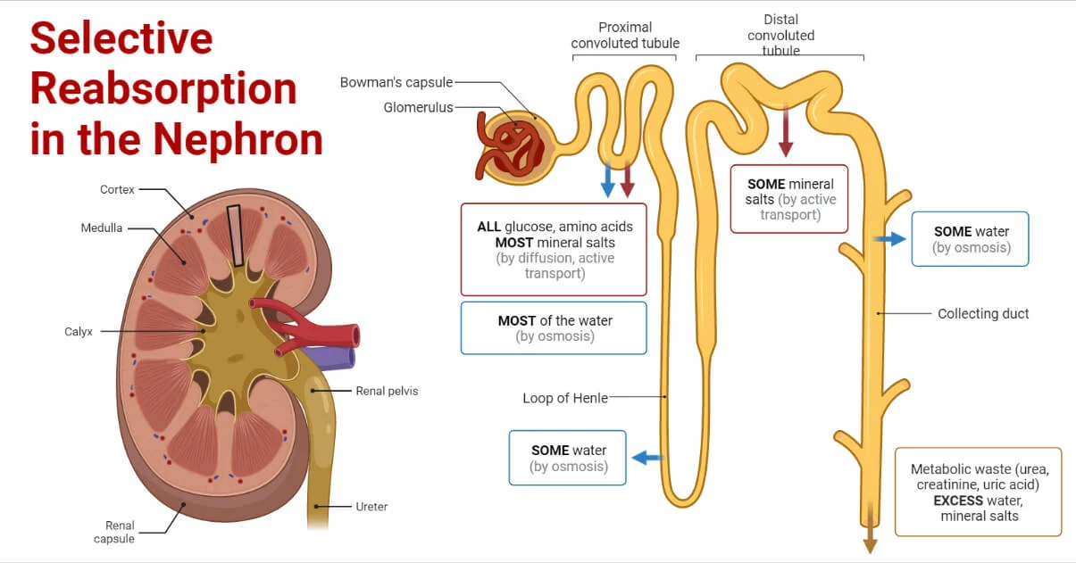
- Ultrafiltration (Bowman's capsule/glomerulus): High blood pressure in glomerular capillaries forces small molecules (water, glucose, amino acids, urea, salts, creatinine) through the filtration membrane into Bowman's space.
- Selective reabsorption (PCT, loop of Henle, DCT): Useful molecules reabsorbed back into blood.
- Tubular secretion (DCT and collecting duct): H⁺ ions, NH₄⁺, K⁺, some drugs secreted from blood into tubular fluid.
Kidney disease in Nigeria: Hypertension and diabetes mellitus are leading causes of chronic kidney disease.
Nervous System I — Neuron, Impulse & Synapse
Neuron structure · Resting potential · Action potential · Synaptic transmission · Reflex arc
Learning Outcomes
- Describe the structure and types of neurons
- Explain the resting membrane potential and how an action potential is generated
- Describe synaptic transmission including the role of neurotransmitters
- Trace the pathway of a reflex arc and give an example
11.1 Neuron Structure
11.2 Action Potential
| Phase | Ion Movement | Membrane Potential |
|---|---|---|
| Resting potential | K⁺ inside; Na⁺ outside (maintained by Na⁺/K⁺ ATPase pump) | −70 mV (inside negative) |
| Depolarisation | Voltage-gated Na⁺ channels open; Na⁺ rushes INTO cell | Rises rapidly to +40 mV |
| Repolarisation | Na⁺ channels close; voltage-gated K⁺ channels open; K⁺ rushes OUT | Falls back toward −70 mV |
| Hyperpolarisation | K⁺ channels slow to close; brief overshoot | Drops slightly below −70 mV |
| Refractory period | Na⁺/K⁺ pump restores ion distribution; Na⁺ channels in absolute refractory state | Returns to −70 mV; no new AP possible briefly |
11.3 Synaptic Transmission & Reflex Arc
- Action potential arrives at pre-synaptic axon terminal
- Voltage-gated Ca²⁺ channels open → Ca²⁺ flows into terminal
- Ca²⁺ triggers synaptic vesicles to fuse with pre-synaptic membrane → exocytosis of neurotransmitter (e.g. acetylcholine)
- Neurotransmitter diffuses across synaptic cleft (~20 nm)
- Binds to receptors on post-synaptic membrane → ion channels open → new action potential (excitatory) or hyperpolarisation (inhibitory)
- Neurotransmitter removed: by enzyme breakdown (acetylcholinesterase breaks ACh) or reuptake into pre-synaptic terminal
- Receptor detects stimulus (e.g. skin pain receptor)
- Sensory (afferent) neuron transmits impulse to spinal cord
- Relay (interneuron) in posterior horn of spinal cord processes and relays
- Motor (efferent) neuron transmits impulse from spinal cord to effector
- Effector (muscle contracts / gland secretes) → response
- Signal also travels up spinal cord to brain — consciousness is aware AFTER the reflex occurs
Nervous System II — CNS, PNS & Sense Organs
Brain regions · Autonomic NS · Eye · Ear · Skin · Taste · Smell
Learning Outcomes
- Name the major brain regions and their functions
- Distinguish sympathetic from parasympathetic nervous systems
- Describe the structure and function of the eye, ear, skin, tongue, and nose as sense organs
12.1 Brain Regions and Functions
| Brain Region | Location | Functions |
|---|---|---|
| Cerebrum (cerebral cortex) | Largest; two hemispheres; anterior | Voluntary movement, thought, memory, language, consciousness, sensory interpretation, personality |
| Cerebellum | Posterior, inferior | Balance, coordination, fine motor control, muscle tone; damage → ataxia |
| Medulla oblongata | Inferior brainstem | Vital centres: respiratory rhythm, heart rate, blood pressure, swallowing, vomiting, coughing; cranial nerves IX–XII |
| Hypothalamus | Floor of diencephalon | Thermoregulation (body temperature set point), hunger/satiety, thirst, sleep-wake cycle, controls pituitary gland (master of endocrine system) |
| Thalamus | Central diencephalon | Sensory relay station — all sensory input (except olfaction) passes through thalamus before reaching cortex |
| Pons | Brainstem, between medulla and midbrain | Relay between cerebrum and cerebellum; breathing centres; cranial nerves V–VIII |
12.2 Sympathetic vs. Parasympathetic
| Feature | Sympathetic | Parasympathetic |
|---|---|---|
| Activation state | Fight-or-flight; exercise; stress; emergency | Rest-and-digest; sleep; recovery |
| Heart rate | ↑ Increases | ↓ Decreases (vagus nerve) |
| Pupils | Dilated (mydriasis) | Constricted (miosis) |
| Bronchioles | Dilated (bronchodilation) — more O₂ | Constricted (bronchoconstriction) |
| Digestive system | Inhibited (peristalsis slows, sphincters close) | Stimulated (peristalsis increases, digestive glands active) |
| Neurotransmitter | Noradrenaline at target organs | Acetylcholine at target organs |
12.3 Sense Organs Summary
| Sense Organ | Stimulus | Receptor Cells | Key Structures / Pathway |
|---|---|---|---|
| Eye | Light (photons 380–750 nm) | Rods (dim light, b&w, rhodopsin); Cones (colour, iodopsin — 3 types) | Cornea (refraction) → aqueous humour → iris/pupil → lens (accommodation by ciliary muscles) → vitreous humour → retina → fovea (highest acuity) → optic nerve → visual cortex |
| Ear | Sound waves; gravity/rotation (vestibular) | Hair cells in Organ of Corti (hearing); hair cells in semicircular canals/utricle/saccule (balance) | Pinna → ear canal → tympanum → 3 ossicles (malleus, incus, stapes) → oval window → cochlea (perilymph → endolymph waves → hair cell bending → impulse) → auditory nerve → auditory cortex |
| Skin | Touch, pressure, temperature, pain, vibration | Meissner's (light touch); Pacinian (deep pressure/vibration); Merkel's (sustained touch); Ruffini (skin stretch); free nerve endings (pain/temperature) | Stimulation → sensory neuron → spinal cord → thalamus → somatosensory cortex |
| Tongue | Chemical (taste = gustation) | Taste receptor cells in taste buds (on papillae) | 5 basic tastes: sweet (sugars), sour (H⁺), salty (Na⁺), bitter (alkaloids), umami (glutamate/protein). 3 cranial nerves carry taste: VII (anterior 2/3), IX (posterior 1/3), X (epiglottis) |
| Nose | Chemical (smell = olfaction) | Olfactory receptor neurons (bipolar neurons) in olfactory epithelium (roof of nasal cavity) | Volatile chemicals → olfactory receptor proteins → olfactory nerve (CN I) → olfactory bulb → olfactory cortex + limbic system (explains strong emotion-smell link) |
Skeletal System & Muscular Contraction
Skeleton functions · Axial/Appendicular · Joints · Sliding filament theory
Learning Outcomes
- State five functions of the mammalian skeleton
- Distinguish axial from appendicular skeleton
- Classify joints and give examples
- Explain the sliding filament theory of muscular contraction step by step
13.1 Functions of the Skeleton
13.2 Sliding Filament Theory of Muscular Contraction
The sliding filament theory (Hanson and Huxley, 1954) explains how sarcomeres shorten during muscle contraction. Actin (thin) filaments slide over myosin (thick) filaments — the filaments themselves do not shorten, but the sarcomere (and thus the muscle) shortens.
Rigor mortis occurs because after death, ATP is depleted — myosin heads remain bound to actin (no ATP to detach cross-bridges) → muscles stiffen. This resolves after ~24–48 hours as muscle proteins degrade.
Endocrine System & Hormones
Pituitary tropic hormones · Thyroxine · Adrenaline · Reproductive hormones · Insulin
Learning Outcomes
- Define hormone and state five properties of hormones
- Name the pituitary hormones, their targets, and effects
- Describe the effects of thyroxine, adrenaline, and insulin/glucagon
- Name the reproductive hormones and their effects
14.1 Properties and Definition of Hormones
A hormone is a chemical messenger secreted by an endocrine gland directly into the bloodstream, transported to a target organ, where it produces a specific physiological effect. The endocrine system and nervous system together maintain homeostasis — the nervous system acts quickly (milliseconds) but briefly; the endocrine system acts slowly (minutes to hours) but with prolonged effects.
- Secreted by specialised endocrine (ductless) glands directly into blood
- Transported in blood to target organs — may travel long distances
- Effective in very small (nanomolar) concentrations
- Specific — act only on cells bearing complementary receptors for that hormone
- Slow-acting but long-lasting compared to nerve impulses
- Chemical nature: protein/peptide (water-soluble, bind surface receptors) OR steroid (lipid-soluble, enter cell and bind nuclear receptors → gene regulation)
14.2 Pituitary Gland — The Master Gland
| Hormone | Target | Effect / Deficiency / Excess |
|---|---|---|
| GH (Growth Hormone) | Liver, bone, muscle | ↑ Protein synthesis, bone growth; deficiency: pituitary dwarfism; excess in childhood: gigantism; excess in adult: acromegaly |
| TSH (Thyroid-Stimulating H.) | Thyroid gland | Stimulates thyroxine synthesis and secretion |
| ACTH (Adrenocorticotropic H.) | Adrenal cortex | Stimulates cortisol secretion; stress response |
| FSH (Follicle-Stimulating H.) | Ovary / Testis | Female: stimulates follicle development and oestrogen secretion. Male: stimulates spermatogenesis |
| LH (Luteinising H.) | Ovary / Testis | Female: triggers ovulation (LH surge), forms corpus luteum. Male: stimulates testosterone secretion from Leydig cells |
| ADH (Antidiuretic H.) | Kidney collecting ducts | ↑ Water reabsorption → concentrated urine; deficiency: diabetes insipidus (large volumes of dilute urine) |
| Oxytocin | Uterus, mammary glands | Uterine contractions during labour; milk ejection reflex (positive feedback) |
| Prolactin | Mammary glands | Stimulates milk production (lactation) after childbirth |
14.3 Key Hormones — Thyroxine, Adrenaline, Insulin, Reproductive
Reproductive System, Fertilisation & Course Synthesis
Male/female anatomy · Fertilisation · Gestation · Birth · Menstrual cycle
Learning Outcomes
- Describe the structure and function of the male and female reproductive systems in mammals
- Trace the process of fertilisation, implantation, and gestation
- Explain the events of childbirth (parturition)
- Describe the hormonal control of the menstrual cycle
15.1 Male Reproductive System
| Structure | Function |
|---|---|
| Testes (in scrotum, ~2°C below body temperature) | Produce spermatozoa (spermatogenesis) and testosterone |
| Epididymis | Site of sperm maturation and storage (~3 weeks) |
| Vas deferens | Transports sperm from epididymis to ejaculatory duct |
| Seminal vesicles | Secrete fructose (sperm energy), prostaglandins, and alkaline fluid (~60% of semen volume) |
| Prostate gland | Secretes alkaline fluid that neutralises vaginal acidity; contains enzymes that liquefy semen |
| Bulbourethral (Cowper's) glands | Pre-ejaculatory lubricating fluid; neutralises residual urine acidity in urethra |
| Urethra / Penis | Common passage for urine and semen; sexual intercourse and ejaculation |
15.2 Female Reproductive System
| Structure | Function |
|---|---|
| Ovaries (two, in pelvic cavity) | Produce ova (oogenesis) and sex hormones (oestrogen, progesterone) |
| Fallopian tubes (oviducts) | Transport ovum from ovary to uterus; site of fertilisation (usually at ampulla) |
| Uterus | Site of implantation and foetal development; muscular wall (myometrium) contracts during birth; inner lining = endometrium (shed during menstruation) |
| Cervix | Lower narrow part of uterus; normally closed; dilates during birth; secretes mucus (thick mid-cycle, watery at ovulation) |
| Vagina | Birth canal; receives penis during intercourse; acidic environment (pH 3.5–4.5) prevents infection |
15.3 Fertilisation, Gestation, and Birth
15.4 The Menstrual Cycle (~28 days)
- Days 1–5 — Menstruation: Corpus luteum degenerates → progesterone and oestrogen fall → endometrium sheds → menstrual flow. FSH begins to rise.
- Days 6–13 — Follicular Phase: FSH stimulates follicle growth in ovary → follicle secretes increasing oestrogen → endometrium rebuilds (proliferative phase). Oestrogen rises sharply by Day 12–13.
- Day 14 — Ovulation: High oestrogen triggers LH surge from anterior pituitary → mature Graafian follicle ruptures → secondary oocyte released into fallopian tube.
- Days 15–28 — Luteal Phase: Ruptured follicle → corpus luteum → secretes progesterone (and oestrogen) → endometrium thickens further (secretory phase, preparing for implantation). If no fertilisation: corpus luteum degenerates Day 24–26 → hormone levels fall → menstruation begins (Day 1 of next cycle).
15.5 Course Synthesis — All 12 Systems Integrated
| System | Key Concept | Clinical Importance (Nigeria) |
|---|---|---|
| Digestive | Hydrolytic enzymes at each site; bile emulsifies fats; absorption via villi | Malnutrition; diarrhoeal disease; food contamination |
| Circulatory | Double circulation; cardiac conduction; ABO groups; clotting cascade | Hypertension; sickle-cell disease; HIV transmission via blood |
| Respiratory | Alveolar exchange; aerobic respiration; 38 ATP per glucose | Asthma; TB; COVID-19 respiratory complications |
| Excretory | Ultrafiltration → selective reabsorption → secretion; deamination → urea | Kidney disease (hypertension + diabetes driven); malaria-related nephrotic syndrome |
| Nervous | Action potential; synaptic transmission; reflex arc; brain regions | Epilepsy (neurocysticercosis); stroke; meningitis |
| Skeletal/Muscular | Sliding filament; levers; red bone marrow; joint types | Rickets (Vit D/Ca deficiency); fractures; sickle-cell bone pain |
| Endocrine | Pituitary tropic hormones; thyroxine; adrenaline; insulin | Goitre (iodine deficiency); Type 2 diabetes (rising epidemic) |
| Reproductive | Male/female anatomy; fertilisation; gestation; menstrual cycle | Maternal mortality; antenatal care; family planning |
You have completed BIO 222 — from enzyme kinetics to childbirth, from the action potential to the Krebs cycle. Structure and function are inseparable. Use this knowledge well in your teaching.